Surgical Theater is Simulating Patient's Anatomy Scans Using AR/VR Technologies

Surgical Theater presented the economic value of PrecisionVR Enterprise at the 10th Annual Becker's Hospital Review Meeting, Chicago, IL in April 2019.
Precision VR, offers an intuitive and immersive way to engage patients and visualize surgical procedures for Neuro, ENT and Spine, Cardiac and Thoracic.
The hospital can deploy its Enterprise Solution to a hospital-wide scale. This includes satellite locations. While Precision VR, can be connected to hospital's network, it must comply with security policies. All 360° VR cases stored at the hospital's datacenter are available to any Precision VR application on the network.
Moty Avisar, CEO and co-founder of Surgical Theater says Surgical Theater's visualization platform transforms healthcare allows surgeons and patients to fully immerse themselves in the patient's anatomy, giving them a unique view of their individual anatomy. The platform has been proven to increase patient satisfaction and economic outcomes.
The 360° virtual reality technology Precision VR, allows patients to see inside their anatomy with their surgeon. Precision VR, which is based on the CT and MRI scans of the patient, creates a 360° virtual tour that the surgeon and patient can enjoy. The surgeon can literally walk with the patient through their own pathology by placing the VR headset on them. This enhances their understanding and helps to plan their treatment. This joint walk-through of the anatomy has been shown to improve patient satisfaction, patient engagement, and hospital financial performance.
Surgeons can literally walk in the space between white matter tracks, tumor, and gain unprecedented situational awareness, clarity, and assessment. The surgeon can examine the anatomy further and plan the craniotomy and surgical path, trajectory and corridor.
Surgical Theater Introduces a New Approach Using Augmented Reality with Magic Leap
In April 2019 was the debut of AR by Surgical Theater. This AR combined Precision VR, patient-specific models, and the Magic Leap One headset at the American Association of Neurological Surgeons Annual Conference in San Diego, CA, which featured AR virtual reconstructions.
Patients and surgeons are able to walk together for the first time ever in Neurosurgery. Neurosurgical consultations can be facilitated by the 360° AR shared approach. This creates an environment that encourages collaboration and decision making. Augmented Reality allows surgeons to maintain awareness and eye contact with each other while navigating through the virtual 360° model.
By combining Surgical Theater’s Precision VR, reconstructions and data from the patient's charts viewed in AR with the Magic Leap may lead to a limitless number of possibilities. This could include seamless workflows, improved efficiency, transparency in charting, patient satisfaction, and increased efficiency.
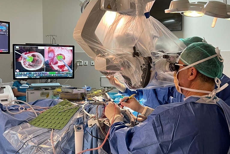
Surgical Theater also showcased SynchronizAR, a Synchronized AR Visualization Platform for Neurosurgery that aligns patient-specific Precision VR, reconstructions with the most advanced endoscope and navigation systems. SynchronizAR allows surgeons to move beyond the hand drawn pathology outlines as seen in standard surgical navigations systems. It facilitates comprehensive MR by offering surgeons live and coordinated augmentation, including pathology and vascular structures.
Moty Avisar believes that Surgical Theater's evolution into MR will allow them to use their core competencies in AR and VR. In the OR, overlaying the 360° model onto the surgeons microscopic/endoscopic optics, enables them to see and therefore, preserve a hidden vessel or nerve that will be exposed during a tumor resection. Surgical Theater hospital partners have found the 360° model to be extremely valuable in their clinics. It has helped improve patient understanding and resulted in patient satisfaction.
There are many medical institutions across the world that use this technology, including George Washington University Hospital and UCSF Benioff children's hospital, Stanford Medical Center, Houston Methodist Hospital, Stanford Medical Center, Stanford Medical Center, Stanford Medical Center, Houston Methodist Hospital, NYU, University Hospitals and Mt. Hoag Hospital and Sinai use Precision VR(r), a surgical theater's virtual reality system, in both the operating room and in the clinic.
A retrospective review of patients was done from the 9-months prior to VR implementation up to the 9-months after. The patient retention rate went up from 64% to 96%, a 50% increase. The outmigration rate dropped from 36% down to 4%, an 89% decrease. The result was that the contribution margin from Precision VR, which saw 113% increase in implementation, significantly increased. These results demonstrate how Precision VR(r can be used across the patient care continuum, and how it has been economically successful.
Surgical Theater Responds To COVID-19 Pandemic With Free Remote 360° VR Services
Surgical Theater provided its VR technology for free to all hospitals that wanted to enter the lungs of COVID-19 patients in June 23, 2020.
Surgical Theater used patient-specific MRIs and CT scans to create a 360° VR environment. This allows doctors to "walk" and navigate through the patient's anatomy. Medical teams can better understand the severity of each patient's disease with this FDA Cleared and CE Mark technology. For patients with COVID-19, Precision VR, which can be used to identify lesions that are specific to the virus, will help distinguish them from other forms of lung damage and chronic pulmonary disease. This will help to understand the pre-existing condition patients who are most at risk. Visual representations of COVID-19-specific lesions in volume-representation could be used to enhance the use of chest CT images. This will allow for better understanding of disease progression, resolution and long-term effects. Volume measurements of the affected tissue in the lungs could be derived from patient scans and greatly improve treatment algorithms for this pandemic.
Many COVID-19 patients get pneumonia. This can lead to respiratory failure, and even death. The symptoms of COVID-19 pneumonia are different to other forms of bacterial pneumonia. These differences can be seen in chest CT scans. The most striking features are the cloudy lesions, which look like shards or reticular lines. These opaque lesions appear to be irregular paving tiles and surround the peripheries. The majority of bacterial pneumonia lesions are found in one lung, and they may not look like shards or glass.
The hospital uploads CT scan results to Surgical Theater's servers. This will create a 3D immersion image of patient's lungs.
Moty Avisar stated that Surgical Theater recognizes and supports the urgent need for precise imaging tools to save lives in the treatment of COVID-19. A global response was needed to this global crisis. Many doctors are still unaware of this and the company sought to help hospitals better understand COVID-19 and provide tools for them to treat it. The Surgical Theater technology has been used by the top American medical institutions to combat COVID-19.
The Surgical Theater 360XR Visualization was used to successfully separate extremely rare conjoined twins in Israel
A rare operation that was only 20 times in the world was performed in Israel on September 20th, 2021 using Surgical Theater's 360XR Visionization.
Although surgery is a daunting task in itself, it is not something that parents can imagine. All the uncertainties, all fears, and all the potential outcomes that can't be predicted. The medical staff at Soroka Medical Center had access to the most advanced medical applications that could help reduce the risks associated with complex surgeries. One of these technologies was developed by Surgical Theater in Israel, an American company. It played a crucial role in giving the medical team tools that were beyond their visual limits.
This complex procedure involved twins who were fused at the head. It took a team at Soroka Medical Center, Israel. One-year-old partially craniopagus half-siblings were involved. They had been attached at the back of each other's heads and facing one another. The Surgical Theater 360deg VR models offered advanced insight to analyze blood vessels and plan for separation.
This is an extremely complex, high-risk operation that can be dangerous and complicated. It was performed in Israel for the first time.
The 360° VR rendered model was reviewed by the surgeons during preoperative surgical planning. They were able see the major veins around each patient's brain surface. The superior sagittal sinus, a major vein that brings venous blood back to the heart from the brain, was found in the exact center of the place where the twins fused. This fact was known by surgeons before using Surgical Theater's virtual modeling. However, it was not possible to determine the junction points and paths of the smaller but still vital veins that lie within the fusion area. The 360XR model allowed surgeons to accurately analyze and understand the location and function of the shared vein that runs between the right transverse and superior sagittal sinuses of Patients 1 and 2. Virtual modeling enabled for a better understanding of the abnormality of the right transverse Sinus of Patient 2. Virtual imaging provided surgeons with the best tool to visualize anatomy. This was something that 2D CT imaging alone could not provide.
Surgical Theater's VR model allowed for accurate simulation of the analysis and anatomy required for the operation. This system allowed surgeons to better understand the surgical field and examine the relationships between them. It also helped them plan their operations in the most efficient way.
Surgical Theater's 360° VR system continues to be a valuable tool for surgeons when paired with intraoperative navigation. Integrated navigation allowed surgeons to utilize their greater knowledge of anatomy from start to finish. Virtual mapping allowed surgeons to track the exact location of the incisions and craniotomies made during preoperative planning. It also allowed them to avoid and see blood vessels.

